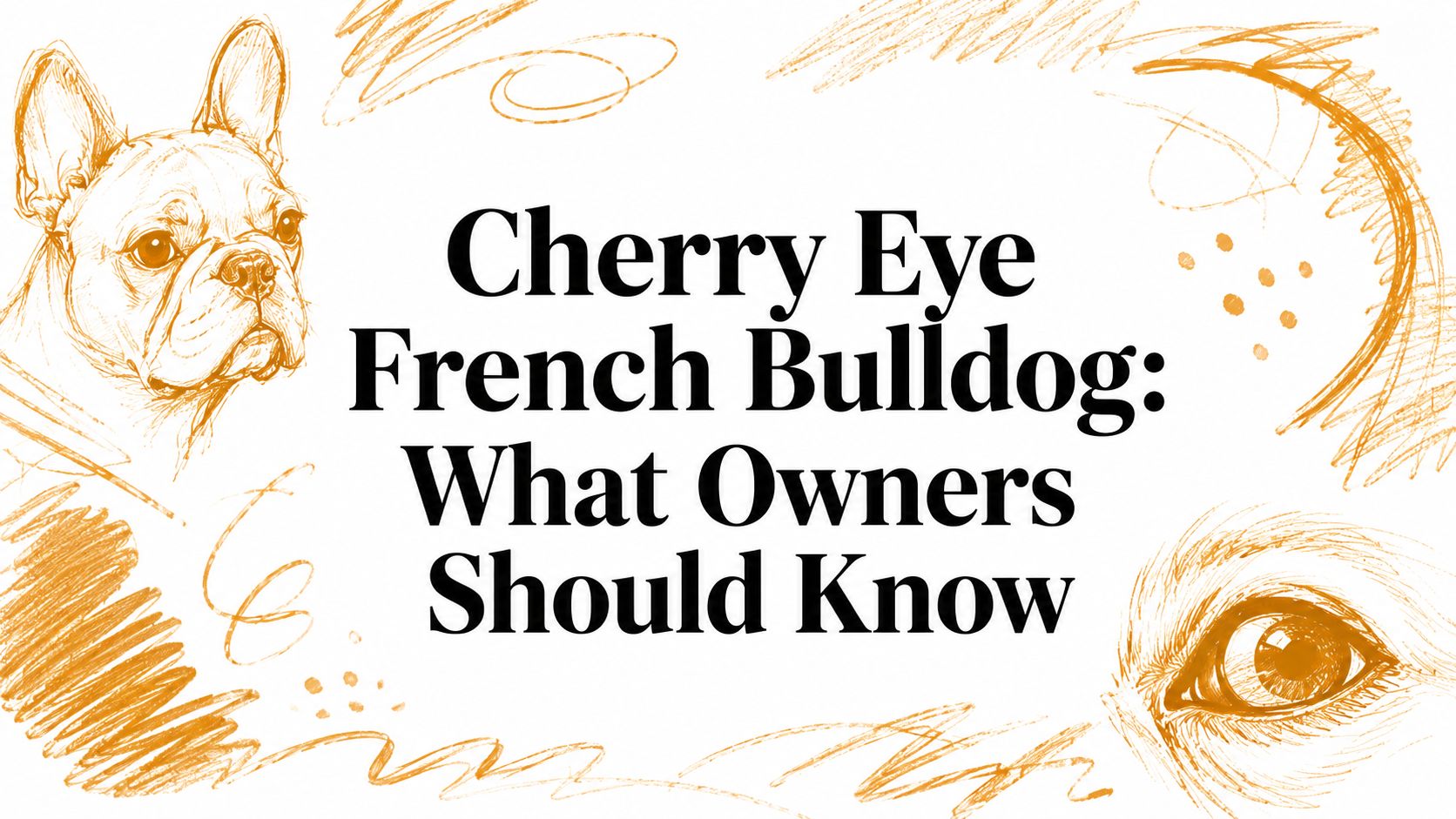
You're cuddling your French Bulldog when you notice a smooth, pink-red lump at the inner corner of one eye. Your dog may seem mostly comfortable, but the appearance is alarming. You check for discharge, pawing, or a visible scratch, then wonder whether the mass is an injury, an infection, or something more serious.
That first reaction is understandable. In a French Bulldog, the finding may be cherry eye, a prolapse of the gland attached to the third eyelid. It usually needs veterinary attention, but the visible bulge is only the beginning of the decision. Owners also need to understand gland preservation, recurrence, the possibility of disease in the other eye, and the broader eye problems associated with brachycephalic anatomy.
Table of Contents
- Spotting the Red Mass in Your French Bulldog's Eye
- What Cherry Eye Is and Why French Bulldogs Get It
- How Vets Confirm the Diagnosis
- Treatment Options From Conservative Care to Surgery
- Recurrence, the Other Eye, and Long-Term Outlook
- What Treatment Typically Costs Across the United States
- How Pet Insurance Handles Cherry Eye Claims
- Your Next Steps and Questions for the Vet
Spotting the Red Mass in Your French Bulldog's Eye
Maya's owner noticed the change during an evening cuddle. One eye looked normal, while the other had a rounded, pink-red swelling tucked into the inside corner. Maya wasn't crying, but she blinked more often and rubbed her face once against the sofa. Her owner's first thought was a scratch. The second was a tumor.
A smooth mass in that location is often consistent with cherry eye, but owners can't confirm the cause from appearance alone. The third eyelid can become visible for other reasons, including sedation, dehydration, or weight loss, as AAHA's overview of the third eyelid explains. A corneal injury, inflammation, prolapsed cartilage, or another eye disorder can also produce an abnormal appearance.
What to do during the first hour
Start by documenting what you see without touching the eye.
- Take a clear photograph: Use good light and capture the affected eye from the front and side. A photo can help the veterinary team judge whether the swelling changes before the appointment.
- Compare both eyes: Note whether the opposite eye looks normal, watery, swollen, or partly covered by the third eyelid.
- Watch behavior: Record blinking, squinting, face rubbing, discharge, sensitivity to light, and changes in appetite or activity.
- Avoid pressure: Don't press, squeeze, scrape, or repeatedly pull the eyelids open. Manipulation can increase irritation and may worsen an injury.
- Call your veterinarian: Describe the color, location, onset, and any signs of discomfort when you contact the clinic.
The French Bulldog breed health reference can provide broader context, but a new eye mass still deserves an individual examination. Seek urgent veterinary guidance if your dog is squinting continuously, keeping the eye shut, showing sudden swelling, or producing thick discharge. Those signs can indicate painful surface disease rather than an uncomplicated gland prolapse.
What Cherry Eye Is and Why French Bulldogs Get It
A young French Bulldog suddenly develops a smooth pink lump at the inner corner of one eye. That lump is often called cherry eye, but it is not a new growth. It is usually the tear-producing gland of the third eyelid, also called the nictitating membrane, slipping out of position.
The third eyelid works like a small protective panel and lubricating system. Its gland helps spread and produce tears across the eye's surface. Fibrous tissue normally anchors the gland, much like a bracket holding a shock absorber in place. If that support is weak or loose, the gland can shift forward and become visible.
French Bulldogs have a brachycephalic, or flat-faced, skull shape. Their facial structure is associated with prominent eyes, altered eyelid support, and a higher likelihood of several ocular problems. Veterinary epidemiology identifies French Bulldogs among breeds predisposed to cherry eye. The RVC VetCompass cherry-eye infographic reports that flat-faced breeds had up to 34 times the risk of medium-skull dogs.

Why age and recurrence matter
Cherry eye commonly appears early in life. A 2024 peer-reviewed genomic study reported a lifetime prevalence of 0.88% in dogs overall, with most diagnoses made during the first year, as summarized in this French Bulldog cherry-eye evidence review. The same review notes an annual overall prevalence of 0.20% in RVC VetCompass data and a 6.9-times higher risk for brachycephalic breeds than for medium-length-skull breeds.
These figures describe dogs overall and breed groups, not one French Bulldog's personal forecast. A young dog with cherry eye may later develop the condition in the other eye or experience recurrence after treatment. That changes the lifetime cost calculation: owners may need to plan for more than one examination or procedure, while insurance may assess each eye, recurrence, and policy exclusions separately. The Bulldog breed health page provides related breed context, but an eye examination determines what is happening in an individual dog.
How Vets Confirm the Diagnosis
A veterinarian usually begins with history, not instruments. The clinic will want to know when the swelling appeared, whether it has changed size, whether your dog is rubbing or squinting, and whether there has been discharge or recent trauma. Bring the photograph you took at home, especially if the gland moved back temporarily.
The visual examination then separates a likely gland prolapse from other causes of a visible third eyelid. The vet assesses the tissue's position and color, looks for inflammation, checks the cornea, and compares the two eyes. A red mass at the inner corner supports cherry eye, but it doesn't prove that the gland is the only problem.
What the appointment may include
| Exam Step | What It Checks | Why It Matters |
|---|---|---|
| History and observation | Onset, rubbing, squinting, discharge, and changes in behavior | Helps establish urgency and distinguish irritation from a sudden painful injury |
| Eyelid and third-eyelid examination | Gland position, swelling, cartilage shape, and surrounding tissue | Helps separate cherry eye from prolapsed cartilage or another mass |
| Schirmer tear test | Tear production in each eye | Establishes whether the eye is producing adequate tears and provides a baseline for follow-up |
| Fluorescein stain | Corneal scratches or ulcers | Identifies surface damage that may require faster treatment |
| Eye comparison | Differences between the affected and unaffected sides | Can reveal early disease in the other eye or broader facial-structure problems |
A confident general-practice diagnosis may be appropriate when the gland is clearly prolapsed and the cornea appears healthy. Referral to a veterinary ophthalmologist becomes more useful when the diagnosis is uncertain, the cornea is damaged, tear production is abnormal, the tissue has returned after repair, or the dog has several brachycephalic eye abnormalities.
Urgent finding: Continuous squinting, a cloudy cornea, sudden swelling, marked pain, or thick discharge should move the visit out of routine scheduling and into urgent veterinary assessment.
The exam also protects owners from treating the wrong condition. A visible third eyelid isn't automatically cherry eye, and a seemingly simple pink bulge can coexist with a painful corneal problem.
Treatment Options From Conservative Care to Surgery
Your French Bulldog's red gland may look like a small surface problem, but treatment must protect the eye now and preserve tear production later. The plan depends on the gland's condition, the eye surface, your dog's comfort, and anesthesia risk. Conservative care can reduce irritation or briefly return a newly prolapsed gland to position. It usually does not repair the weak attachment that allowed the gland to slip.
The gland works like a small anchor that has come loose from its mooring. Lubricating medication may protect exposed tissue while you arrange an appointment. A veterinarian might also prescribe anti-inflammatory treatment or demonstrate a gentle manual reduction for a selected early case. Warm compresses may soothe nearby tissue when the veterinary team approves them. Forceful massage and human eye drops can cause harm without veterinary direction.
Comparing the realistic choices
| Approach | Potential role | Main limitation |
|---|---|---|
| Lubricating medication | Protects the exposed gland and supports comfort while awaiting care | Does not reliably restore the gland's normal position |
| Warm compresses | May ease local irritation when approved by a veterinarian | Cannot repair the connective-tissue attachment |
| Manual reduction | May temporarily reposition the gland in selected early cases | Prolapse can return, so this is not a dependable cure |
| Gland-sparing surgery | Repositions the gland while preserving its tear-producing function | Requires anesthesia, sutures, aftercare, and possible revision |
| Gland removal | Removes the visible prolapse | Reduces tear-producing tissue and raises long-term dry-eye risk |
The usual goal is surgical gland replacement, not excision. Cornell's cherry-eye guidance explains why preserving the gland matters. Repositioning addresses the visible prolapse while retaining tissue that supports the eye's tears.
Pocket and imbrication techniques place the gland beneath surrounding tissue and hold it with sutures. The surgeon aims to restore its position without blocking normal function. Removal may seem simpler, but losing the gland can contribute to keratoconjunctivitis sicca, commonly called chronic dry eye, later in life. Veterinary guidance for Bulldogs and French Bulldogs supports repositioning rather than removal.

On surgery day, expect a preanesthetic assessment, anesthesia, gland repositioning, and recovery monitoring. Your dog may go home with eye medication, an Elizabethan collar, and instructions to prevent rubbing during healing. Ask how the clinic manages anesthesia in brachycephalic dogs, since airway planning and careful recovery monitoring matter for this breed.
Recurrence, the Other Eye, and Long-Term Outlook
A successful repair can still require long-term monitoring. Veterinary summaries commonly describe recurrence as possible, including roughly 10% to 20% of repairs, while UK guidance often reports about one in ten repairs returning, as discussed in this French Bulldog cherry-eye overview. Technique, gland condition, inflammation, and surgeon experience can all affect the result.
The opposite eye also deserves attention. A French Bulldog with cherry eye in one eye may later develop it in the other because the same tissue vulnerability can affect both sides. Preventive surgery is not automatic. A veterinarian may assess the second gland's position, tear production, inflammation, and the risks of another anesthetic before suggesting treatment.
Why the broader eye picture matters
Repair puts one displaced gland back in place. It does not change the facial structure associated with other brachycephalic eye concerns, such as corneal injury, chronic dry eye, proptosis, entropion, and eyelid abnormalities. Veterinary coverage of French Bulldog health concerns places cherry eye within this wider pattern.
Surgery can correct the prolapse while leaving broader eye-care needs unchanged.
That distinction affects lifetime planning and insurance decisions. Track examinations, tear-production testing, medications, and changes in either eye. A recurrence or second-eye diagnosis may mean another anesthesia and recovery period. A separate corneal or tear-film disorder may require continued treatment, adding costs beyond the first repair.

What Treatment Typically Costs Across the United States
The financial question has no single useful national answer. A French Bulldog receiving unilateral gland-sparing surgery at a general veterinary practice may face a very different bill from a dog treated by a board-certified veterinary ophthalmologist in a major coastal metro area. The amount also depends on whether the invoice includes diagnostics, anesthesia, fluids, medication, follow-up examinations, and an e-collar.
Regional variation matters. Veterinary fees often run higher in major metropolitan and coastal markets than in many Midwest and Southern communities, but the local clinic's service mix can matter just as much. Specialty referral adds expertise and may add facility or consultation charges, while a general practice may offer a more direct route for an uncomplicated case.
The line items that change the estimate
| Cost Driver | Effect on Price |
|---|---|
| One eye versus both eyes | Treating both eyes during one anesthetic event may reduce the combined charge compared with staging separate procedures |
| General practice versus ophthalmology referral | Specialty care may increase the bill while offering advanced eye expertise |
| Geography | Local veterinary labor, facility, and operating costs shape the estimate |
| Preanesthetic testing | Bloodwork and other screening add services before anesthesia |
| Anesthesia and monitoring | Drug choice, airway management, and recovery observation affect the total |
| Medications and protection | Take-home eye medication and an e-collar may appear as separate items |
| Follow-up care | Tear testing, rechecks, and revision evaluation can extend the treatment course |
The bigger planning error is looking only at the first repair. A recurrence, disease in the opposite eye, chronic dry-eye medication, or another brachycephalic eye problem can alter the lifetime financial picture. Treat cherry eye as one possible event within a broader French Bulldog eye-care trajectory, not as an isolated line item.
For a state-adjusted example, review the French Bulldog Texas veterinary estimate. Ask your clinic for an itemized estimate that separates the examination, diagnostics, anesthesia, surgery, medications, and rechecks. That format makes it easier to compare providers without comparing incomplete totals.
How Pet Insurance Handles Cherry Eye Claims
Pet insurance decisions require policy reading before the eye appears abnormal. Accident-and-illness coverage may address hereditary conditions, but eligibility can depend on waiting periods, bilateral-condition language, exclusions, deductibles, reimbursement rules, and whether the insurer considers a previous eye symptom pre-existing.
A French Bulldog's predisposition makes timing especially important. If one eye has already been diagnosed before enrollment, a policy may exclude that eye and may also apply a bilateral-condition clause to the other eye. Some plans treat matching conditions in paired organs as related, while others impose specific limits on when the opposite eye can qualify.
Read the policy through the claim
Keep the veterinary invoice itemized. The claim may separate:
- Exam fees: The consultation and diagnostic assessment.
- Testing: Tear-production measurement, staining, or other eye evaluation.
- Anesthesia and surgery: The gland-replacement procedure and operating-room services.
- Medication: Prescribed drops, ointments, or pain control.
- Rechecks: Follow-up examinations and postoperative monitoring.
Coverage can also differ when a repair is described as cosmetic rather than medically necessary. Ask the insurer how it defines hereditary disease, pre-existing signs, bilateral conditions, recurrence, and revision surgery before you enroll. Don't rely on a sales summary when the full policy wording controls the claim.
The PetCostEngine insurance comparison and breakeven tools can help you examine scenarios involving a first-eye claim, recurrence, or treatment of the other eye. Breakeven isn't a promise that a policy will pay more than premiums. It's a way to compare expected covered claims with the policy's premiums and out-of-pocket structure.
A short pre-purchase checklist
- Waiting period: Confirm the length and which conditions it applies to.
- Hereditary wording: Check whether breed-linked conditions are covered or excluded.
- Bilateral clause: Learn how the policy handles the second eye after a first-eye diagnosis.
- Pre-existing definition: Ask whether earlier redness, discharge, or veterinary notes affect eligibility.
- Payout structure: Review annual, per-condition, and lifetime limits, along with cancellation terms.
Your Next Steps and Questions for the Vet
Start with evidence. Photograph the eye, write down when you first noticed the change, and record squinting, rubbing, discharge, appetite, and activity. Call your regular veterinarian the same day for guidance, even if your dog seems comfortable, because a pink mass can look similar to other eye conditions.
Use urgency as the sorting tool:
- Home observation while awaiting advice: Your dog is comfortable, the eye is open, and there's no obvious trauma, heavy discharge, or cloudiness. Keep the dog from rubbing the area and don't apply unapproved medication.
- Prompt veterinary appointment: The mass persists, the dog blinks more than usual, or the tissue looks inflamed. A clinic can assess the gland and protect the cornea.
- Urgent or emergency evaluation: The dog keeps the eye shut, shows severe pain, develops sudden swelling, has a cloudy or visibly damaged cornea, or suffers an injury. Corneal ulceration can threaten vision and needs veterinary treatment.
Questions to bring to the appointment
Print or save this list on your phone:
- Is this definitely prolapse of the third-eyelid gland?
- Is the cornea scratched or ulcerated?
- Are tear production and the other eye normal?
- Will the procedure preserve the gland?
- Which pocket, imbrication, or other repair technique do you recommend?
- How often do you perform this repair in brachycephalic dogs?
- What recurrence risk do you see with this technique and my dog's tissue?
- Should we monitor the unaffected eye or consider treatment during the same anesthetic event?
- What services are included in the itemized estimate?
- What medications, collar, activity limits, and rechecks will follow surgery?
Surgical timing involves trade-offs. Waiting may allow inflammation and rubbing to continue, while immediate anesthesia may need more careful assessment in a dog with airway or other health concerns. Your veterinarian can balance those factors instead of treating the visible bulge in isolation.
For a broader state-, breed-, and age-based planning view, use the French Bulldog ownership cost calculator. Pair that estimate with your veterinarian's itemized treatment plan and the insurance policy's actual exclusions. The clearest decision frame includes three possibilities, the current eye, recurrence, and a later problem in the other eye or elsewhere in the brachycephalic eye system.
PetCostEngine provides breed- and state-adjusted ownership projections, condition planning, and insurance breakeven comparisons that can help you evaluate those scenarios before or after a cherry-eye diagnosis. Visit PetCostEngine to build a personalized French Bulldog plan and bring clearer questions to your veterinarian.
Estimated lifetime and yearly costs for a French Bulldog, state by state, plus whether pet insurance breaks even for the breed.
See French Bulldog costs →
Is insurance worth it for a French Bulldog? →
Joint supplement → Puzzle toys → No-pull harness →
As an Amazon Associate, PetCostEngine earns from qualifying purchases.